【ベストコレクション】 paranasal sinuses anatomy diagram 290402-What are the 4 paranasal sinuses
All paranasal sinuses drain into nasal cavity; · Anatomy of nose and paranasal sinus 1 Anatomy of Nose and Paranasal SinusByDr Mohammed Faez 2 The NoseThe nose consists of the external nose and the nasal cavity, Both are divided by a septum into right and left halvesMiddle meatus frontal sinus, maxillary sinus, anterior ethmoid;

Sinuses Of Nose Human Anatomy Sinus Diagram Anatomy Of The Royalty Free Cliparts Vectors And Stock Illustration Image
What are the 4 paranasal sinuses
What are the 4 paranasal sinuses- · The maxillary sinuses are located on each side of your nose, near the cheek bones The frontal sinuses are located above the eyes, near your foreheadDownload 584 Sinuses Stock Illustrations, Vectors & Clipart for FREE or amazingly low rates!
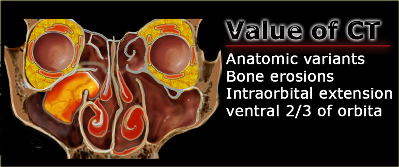


The Radiology Assistant Mri Examination
1604 · Illustration of Human Anatomy Sinus Diagram Anatomy of the Nose Nasal cavity bones Anatomy of paranasal sinuses Sinusitis Antritis It is the inflammation of the maxillary sinuses vector art, clipart and stock vectors ImageEssential for both Medical and non medical students to learn about the basic Anatomy of Nose, Nasal and Paranasal Sinus and their functions Article by Paediatric 99 Respiratory System Anatomy Paranasal Sinuses Maxillary Sinus Internal Carotid Artery Sinus Surgery Nasal Septum Medical Anatomy Dental AnatomyNew users enjoy 60% OFF 165,963,486 stock photos online
· Anatomy of the paranasal sinuses, osteomeatal complex and nasal walls Maxillary sinuses (Fig 1) the maxillary sinus is pyramidal in shape with the apex in the zygomatic process of the maxilla bone and the base at the lateral wall of the nose Behind the posterior wall are the infratemporal and pterygopalatine fossaeFrontal sinus, sphenoidal sinus,;Ethmoid sinus (known as
Anatomy of Paranasal sinuses Abstract Paranasal sinuses are air filled hollow sacs seen around the skull bone These sacs precisely surround the nasal cavity There are four paired sinuses surrounding the nasal cavity This article attempts to trace the history of anatomy of paranasal sinuses from early 16th century till date TheDiagram Of Paranasal Sinuses Download Scientific Diagram The Paranasal Sinuses Structure Function Teachmeanatomy Paranasal Sinus Definition Location Anatomy FunctionLearn more Royaltyfree stock vector ID Nasal sinus Human Anatomy Sinus Diagram Anatomy of the Nose Nasal cavity bones Anatomy of paranasal sinuses Sinusitis Antritis It is the inflammation of the maxillary sinuses l


Paranasal Sinuses Wikipedia


Anatomy And Functions Of The Paranasal Sinuses Youtube
Maxillary Sinus Saved by Alecia Fidler Maxillary Sinus Nose Diagram Palatine Bone Sphenoid Bone Paranasal Sinuses Parts Of The Nose Nasal Septum Molar ToothLearn sinuses paranasal neck anatomy with free interactive flashcards Choose from 500 different sets of sinuses paranasal neck anatomy flashcards on QuizletAnatomy and Physiology of the Nose and Paranasal Sinuses PD Dr med Basile N Landis Unité de RhinologieOlfactologie Service d'OtoRhinoLaryngologie et de Chirurgie cervicofaciale, Hôpitaux Universitaires de Genève, Suisse
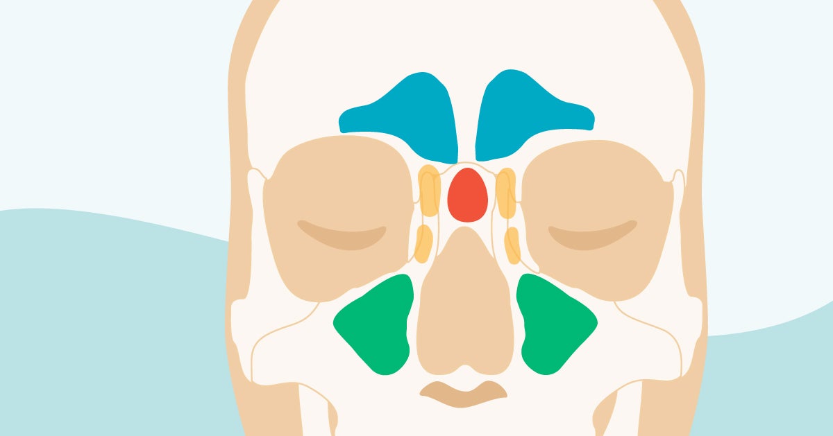


Sinus Cavities In The Head Anatomy Diagram Pictures
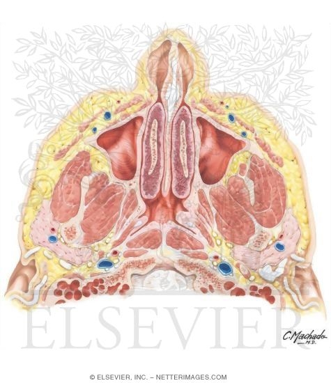


Nose And Paranasal Sinuses Cross Section
Graphical anatomy is discussed for a clear understanding of the nasal cavity and its relationship to adjoining sinuses and danger areas A threedimensional anatomy is complemented with schematic diagrams Developmental Anatomy Nose Development The embryogenesis of the nose and paranasal sinuses is related to the regional embryology of the cranialoralAnatomy and Functions of the Paranasal Sinuses READ MORE BELOW!In this video, we explore the 4 paranasal sinuses and what their proposed functions areINSTAGRAM · Anatomy atlas of the nasal cavity fully labeled illustrations and diagrams of the nose and paranasal sinuses (external nose, nasal cartilages, nasal septum, nasal concha and meatus, bones of the nasal cavity and vessels and nerves)


Paranasal Sinuses Illustration Anatomical Justice


Jaypeedigital Ebook Reader
0019 · Paranasal Sinuses There are four paired paranasal sinuses, one on either side of the midline They develop from ridges in the lateral nasal wall by the eighth week of embryogenesis and continue pneumatization until early adulthood Each sinus isSuperior meatus posterior ethmoid, sphenoid sinus;The paranasal sinuses (latin sinus paranasales) are four bilateral airfilled spaces within bones of the skull surrounding the nasal cavityFour bones of the skull each accommodates a pair of paranasal sinuses that are named according to the bone in which they are locatedThe four sinuses are maxillary sinus,;
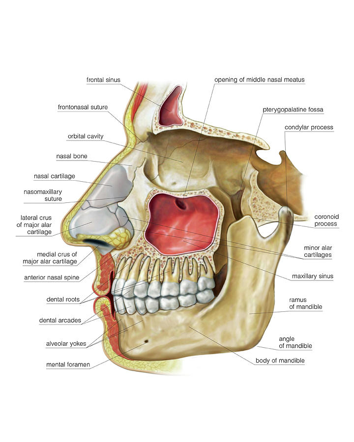


Paranasal Sinuses Photograph By Asklepios Medical Atlas
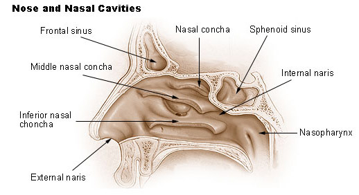


Seer Training Nose Nasal Cavities Paranasal Sinuses
The Anatomy of the Nose The nose is the part of the respiratory tract that sits front and center on your face You use it to breathe air in and to stop and smell the roses The nose's exterior anatomy includes the nasal cavity, paranasal sinuses, nerves, blood supply, and lymphatics2403 · The 4 paranasal sinuses include The frontal, maxillary, ethmoidal, and sphenoid sinuses Each of these sinuses have ducts that drain into the naso/oropharynx Frontal sinuses These are found between the outer and inner aspects of the frontal bone posterior to the superciliary arches, they are funnel shaped and they drain into the nasal cavity via theNose anatomy medical vector illustration diagram with nasal cavity, mouth, sinuses and nose cartilage paranasal sinus stock illustrations Cardiogram Cardiogram, normal heart rhythm on an abstract background paranasal sinus stock illustrations


A Ct Scan In Coronal Plane Shows The Normal Paranasal Sinus Anatomy Download Scientific Diagram


Sphenoidal Sinus Photos And Premium High Res Pictures Getty Images
Semilunar hiatus contains openings to first three, bordered by Uncinate process (anterior/inferior) Ethmoid bulla (posterior/superior) Inferior meatus nasolacrimal duct;Variant anatomy The paranasal sinuses are subject to marked variation between individuals and between sides in the same individual, regarding size (aeration) and bony septations Total paranasal sinus agenesis is very rare 1 Isolated frontal sinus2803 · Ecr 17 C 2117 Ct Anatomy Of Paranasal Sinuses Epos Cerebrospinal Fluid Vector Illustration Anatomical Labeled Diagram With Human Superior Sigittal Sinus Arachnoid Villi Subarachnoid And Spinal Cord


Paranasal Air Sinuses Location Functions Relations And Applied Anatomy Qa


Medical Infographic Of Sinus And Human Nasal Anatomy Medical Infographic Of Sinus Normal Sinus And Sinus Infection Or Canstock
Sinus in anatomy a hollow cavity recess or pocket In bones behind your nose are your sphenoid sinuses There are four pairs of paranasal sinuses the frontal sinuses are located above the eyes in the forehead bone The sphenoid sinuses are Anatomy of the sinusesImaging the paranasal sinuses is routine in clinical practice to evaluate for various sinus pathology, nonspecific facial pain, and preoperative planning for functional endoscopic sinus surgery (FESS), including postoperative followup Our goal is to review the complex sinonasal anatomy, anatomic variants, mucociliary drainage pathways andSinus Anatomy – Paranasal Sinuses Maxillary Sinuses These are found directly in behind the check Ethmoid Sinuses These are smaller pockets of air, often described as having the appearance of honeycomb Frontal Sinuses Your upper uppermost sinuses These are placed directly behind your fore head
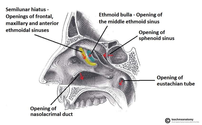


The Paranasal Sinuses Structure Function Teachmeanatomy


1
Anatomy of nose and paranasal sinus This sinus is located inside the face around the area of the bridge of the nose The noses exterior anatomy includes the nasal cavity paranasal sinuses nerves blood supply and lymphatics The bony lateral wall is convoluted by the turbinates called superior middle and inferior turbinate151 MB 3D printed upper respiratory airways cast jpg 1,9 × 2,560; · 1 ANATOMY OF NOSE AND PARANASAL SINUSES DEPT OF OTORHINOLARYNGOLOGY PI M S 2 NOSE ANATOMY DEVELOPMENT Nose develops from frontonasal process which grows between primitive forebrain and the stomodaeum stomodaeum Frontonasal process gets divided into median nasal process and two lateral process Primitive


1


Nasal Sinus Anatomy Anatomy Drawing Diagram
Illustration about pharynx, headache, anatomic, diagram, breathing, sinusitis, organ, system, illness, cavity, head, paranasal, sinus, bone, person, face Illustration of pharynx Stock Photos · #2 Anatomy of the Paranasal Sinuses This excellent 3D model uploaded b y valchanov shows Maxillary sinus one sinus located within the bone of each cheek Ethmoid sinus located under the bone of the inside corner of each eye, although this is often shown as a single sinus in diagrams, this is really a honeycomblike structure of 612 small sinuses that is · Paranasal sinuses in the Horse Sinuses are recesses that arose of outpocketing of nasal canal They are spaces within the bones of the head and aligned with mucosal membrane Not sure of function but probably help decrease weight of skull Horse maxillary sinus is divided into rostral/caudal parts via bony septum, 5 cm behind rostral border of
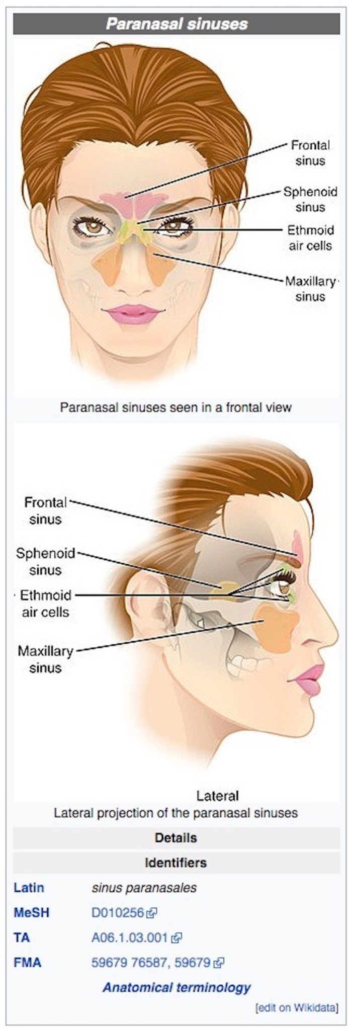


Nathan Lents Is Back Still Wrong About Sinuses Evolution News


Comparison Of Human And Rabbit Sinus Anatomy Download Scientific Diagram
Find Sinuses Nose Human Anatomy Sinus Diagram stock images in HD and millions of other royaltyfree stock photos, illustrations and vectors in the collection Thousands of new, highquality pictures added every day · An interactive quiz covering the Paranasal Sinuses of the Nasal Cavity through multiplechoice questions and featuring the iconic GBS illustrationsMedia in category "Paranasal sinuses" The following 35 files are in this category, out of 35 total 3D printed upper respiratory airways cast jpg 2,560 × 1,9;


Http Www Equisan Com Images Pdf Paranasal Pdf


Paranasal Sinuses
In bones behind your nose are your sphenoid sinuses They're lined with soft, pink tissue called mucosa Normally, the sinuses are empty except for a thin layer of mucusSinus Skull Anatomy · A threedimensional anatomy is complemented with schematic diagrams Developmental Anatomy Nose Development The embryogenesis of the nose and paranasal sinuses is related to the regional embryology of the cranialoralfacial region These primary events occur between the fourth and eighth weeks of fetal life


The Radiology Assistant Mri Examination
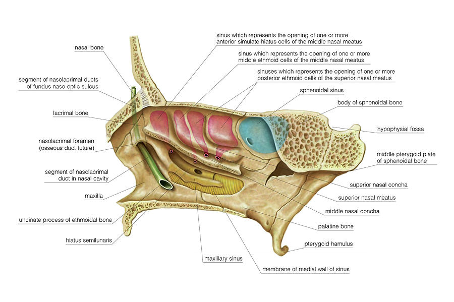


Paranasal Sinuses Photograph By Asklepios Medical Atlas
2804 · Paranasal Sinuses Features Paranasal sinuses are airfilled spaces present within some bones around the nasal cavities The sinuses are Frontal, maxillary, sphenoidal, and ethmoidal All of them open into the nasal cavity through its lateral wall The function of the sinuses is to mack the skull lighter and add resonance to the voice In infections of the sinuses or sinusitis · What are the Paranasal Sinuses Airfilled cavities located within specific facial and skull bones are known as paranasal sinuses 1 Humans have four paired paranasal sinuses, frontal, maxillary, sphenoid, and ethmoid, all extending from the respiratory area of the nasal cavity 2, and named after the bones they are found in Paranasal Sinuses0513 · The paranasal sinuses are airfilled extensions of the nasal cavity There are four paired sinuses – named according to the bone in which they are located – maxillary, frontal, sphenoid and ethmoid Each sinus is lined by a ciliated pseudostratified epithelium, interspersed with mucussecreting goblet cells
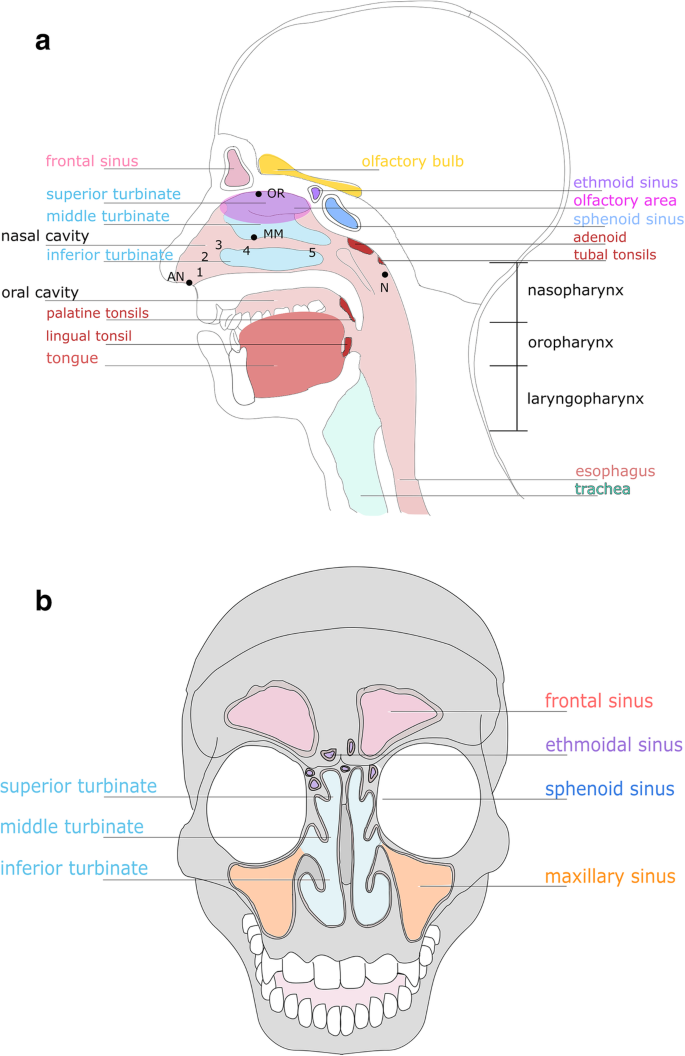


The Microbiome Of The Upper Respiratory Tract In Health And Disease Bmc Biology Full Text


Nasal Anatomical Model Anatomy Of Nose And Paranasal Sinuses Of Frontal Sinus Turbinate Mucosa Artery Olfactory Neural Model Aliexpress
· The ducts and ostia, covered by the nasal turbinates, form the entrances to the paranasal sinuses The size and position of the lateral nasal wall vary widely, dictated by the paranasal sinuses The following endoscopic views should be mentally projected onto the complex of the lateral nasal wallSinuses anatomy paranasal Flashcards Browse 500 sets of sinuses anatomy paranasal flashcardsRegarding blood supply of the paranasal sinuses and the ear The ear is supplied by the posterior auricular and superficial temporal arteries of the external carotid artery The paranasal sinuses are supplied by various branches of the internal and external carotid artery including the maxillary artery, anterior and posterior ethmoidal arteries, ophthalmic artery, supraorbital and
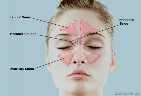


What Are The Sinuses Pictures Of Nasal Cavities
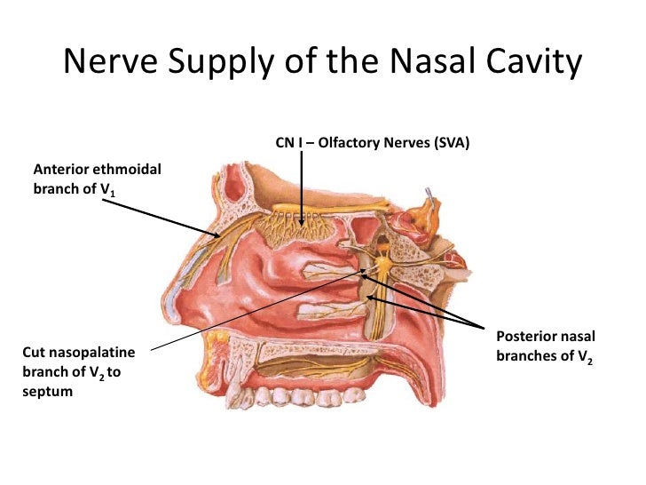


Anatomy Of Nasal Cavity Anatomy Drawing Diagram
Paranasal Sinuses Paranasal sinuses refer to a group of airfilled spaces around the nasal cavity (a system of air channels that connect the nose with the back of the throat) (1) They facilitate the circulation of the air breathed in and out of the respiratory system (2) · The anatomy of the nasal and paranasal cavities supports the numerous functions involved in the protection of the lower respiratory tract The nose and paranasal sinusesImage result for paranasal sinus Saved by LearnAnatomy 13 Gross Anatomy Human Anatomy Nose Diagram Paranasal Sinuses Maxillary Sinus Nose Reshaping Facial Anatomy Nasal Cavity Sinus Problems


The Nasal Cavity And Paranasal Sinuses Canadian Cancer Society


Nose And Paranasal Sinuses Structure And Function Organization Of The Respiratory System
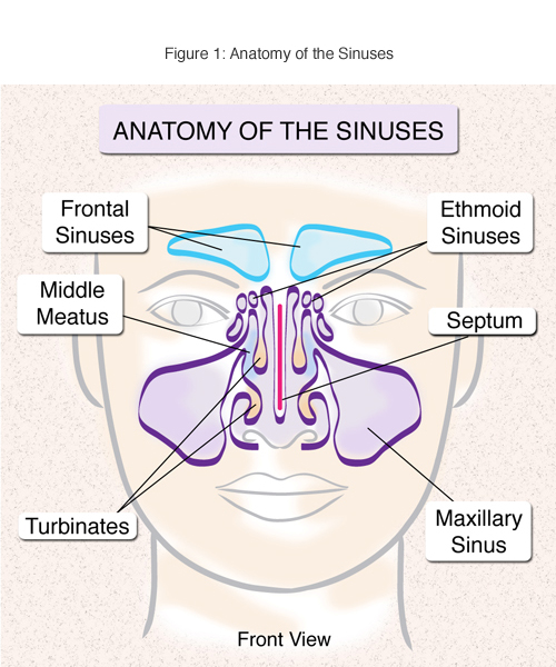


Sinusitis Treatment And Surgery Nyc
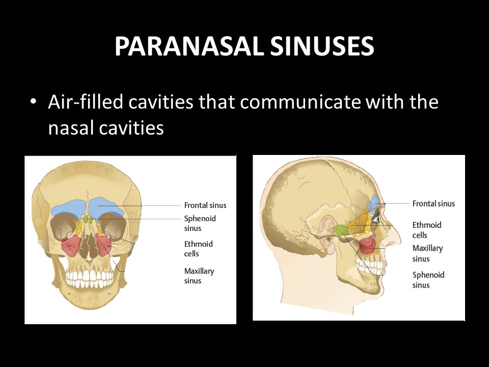


Paranasal Sinuses Anatomy Physiology And Diseases Ppt Video Online Download


Paranasal Sinuses And Nose Anatomy


1
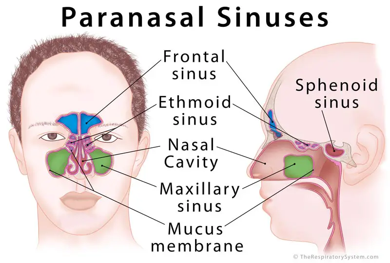


Paranasal Sinus Definition Location Anatomy Function Picture


Human Sinusitis Inflammation Illustration Vector Art Vector Image By C Pattarawit Vector Stock


Ethmoid Sinus Ethmoid Bulla Paranasal Sinuses Anatomy Nose People Anatomy Png Pngegg


Which Of The Following Bones Does Not House A Paranasal Sinus A Ethmoid Bone B Maxillary Bone C Sphenoid Bone D Nasal Bone Study Com
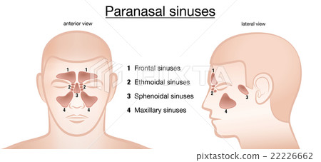


Paranasal Sinuses Anterior Lateral Stock Illustration


Radiology Of The Nasal Cavity And Paranasal Sinuses Ento Key


Figure 3 From Effect Of Functional Endoscopic Sinus Surgery To The Flow Behavior In Nasal During Resting Breathing Condition Semantic Scholar
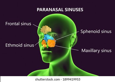


Sphenoid Sinus Images Stock Photos Vectors Shutterstock


1


Sinuses Of Nose Human Anatomy Sinus Diagram Anatomy Of The Royalty Free Cliparts Vectors And Stock Illustration Image


The World S First Precise Model Of The Human Paranasal Sinus For Ess Training 03 03 27


Nasal Cavity Nose Atlas Of Anatomy


Nose Useful Notes On Human Nose And Para Nasal Sinuses Human Anatomy


Nose And Sinus Anatomy Dr Thomas S Higgins Md Msph


Nose And Sinuses Amboss


Ear Paranasal Sinuses Pituitary Gland Bone Anatomy Studying Hard Face Head Human Png Pngwing


7 Paranasal Sinus Illustrations Clip Art Istock


Paranasal Sinuses Illustration Stock Image C025 64 Science Photo Library


Nose And Paranasal Sinuses Structure And Function Organization Of The Respiratory System


Nasal Cavity Paranasal Sinuses Respiratory System Human Mouth Oral Cavity Label People Png Pngegg


Paranasal Sinuses Diagram Quizlet
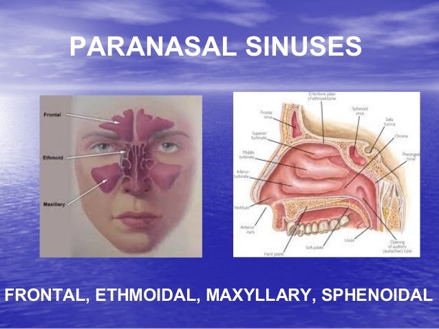


Anatomy Physiology Diseases Of Nose Paranasal Sinuses


Paranasal Sinuses Illustration Stock Image C025 6484 Science Photo Library


Paranasal Sinuses Radiology Key
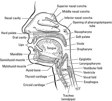


The Anatomy Of The Nose Dummies


Ethmoiditis Images Illustrations Vectors Free Bigstock
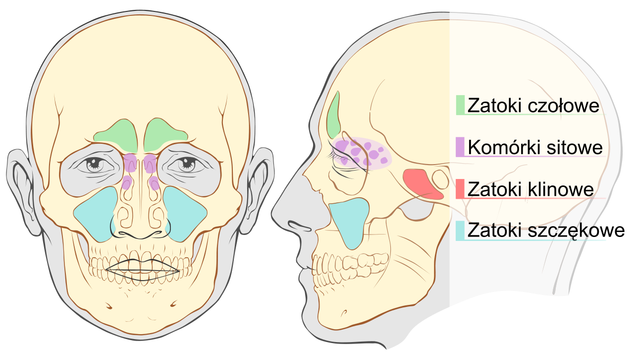


File Paranasal Sinuses Svg Wikimedia Commons


Paranasal Sinuses Wikipedia
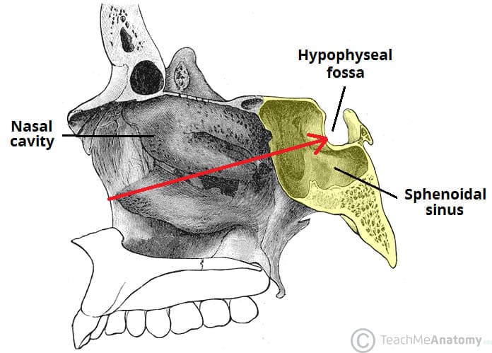


The Paranasal Sinuses Structure Function Teachmeanatomy


Pin On Human Anatomy


Paranasal Sinuses Diagram Quizlet


Maxillary Sinus Radiology Reference Article Radiopaedia Org
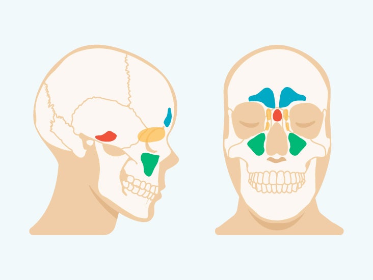


Sinus Cavities In The Head Anatomy Diagram Pictures


Paranasal Sinuses And Sinusitis


Figure Paranasal Sinuses Frontal Sinus Ethmoid Statpearls Ncbi Bookshelf


Pin On Health Tips


Paranasal Sinuses Diagram Quizlet


Surgical Anatomy Of The Paranasal Sinus Ento Key


Facial Sinuses Anatomy Anatomy Drawing Diagram
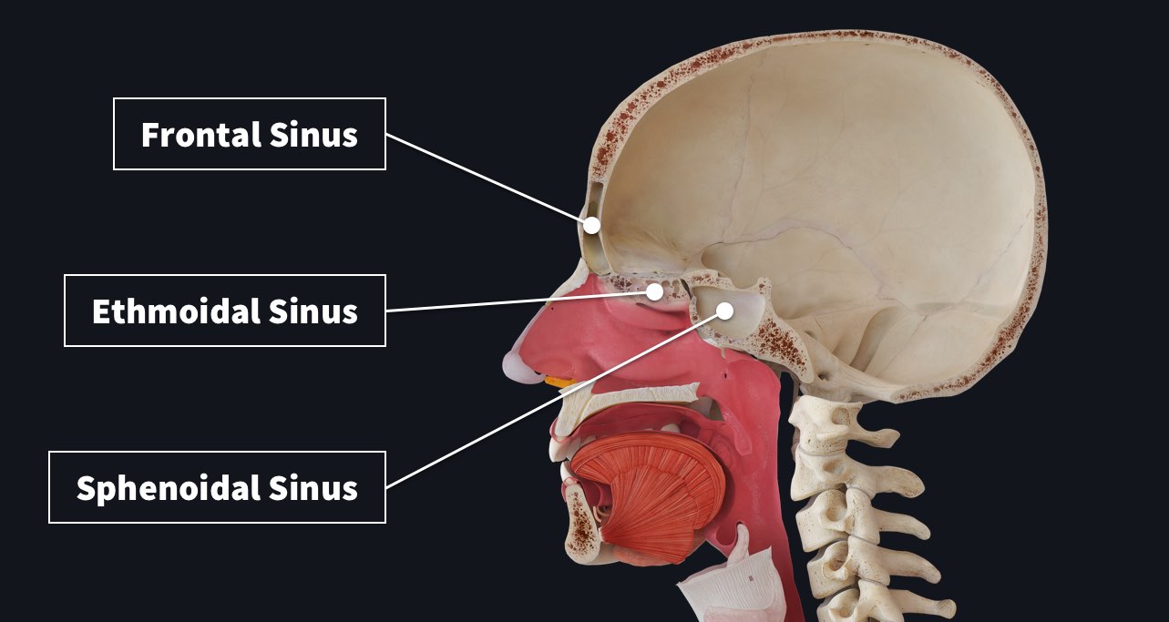


Paranasal Sinuses Complete Anatomy


Paranasal Sinuses Parts Included Frontal Sphenoid Maxillary Sinus Ethmoid Air Cells And Eye Sockets Stock Vector Vector And Low Budget Royalty Free Image Pic Esy Agefotostock


Pin On Nursing School


Introduction To Nose And Sinus Disorders Ear Nose And Throat Disorders Msd Manual Consumer Version


Anatomy Of The Paranasal Sinuses Youtube
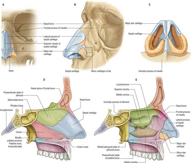


Nose Nasal Cavity And Paranasal Sinuses Clinical Gate


Sinus Model 2850 Anatomy Of Human Sinuses Gpi Anatomicals
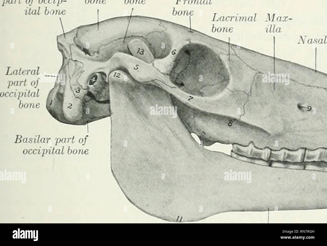


The Anatomy Of The Domestic Animals Veterinary Anatomy The Paranasal Sinuses 85 Superior Maxillary Sinus Is Also Crossed Bj The Infraorbital Canal Over Which It Opens Freeh Into The Sphenopalatine Sinus
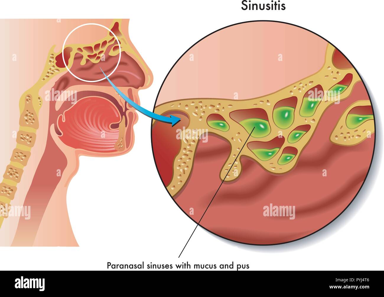


Sinusitis High Resolution Stock Photography And Images Alamy


Normal Anatomy Of The Nose And Paranasal Sinuses Mi Tec Medical Publishing
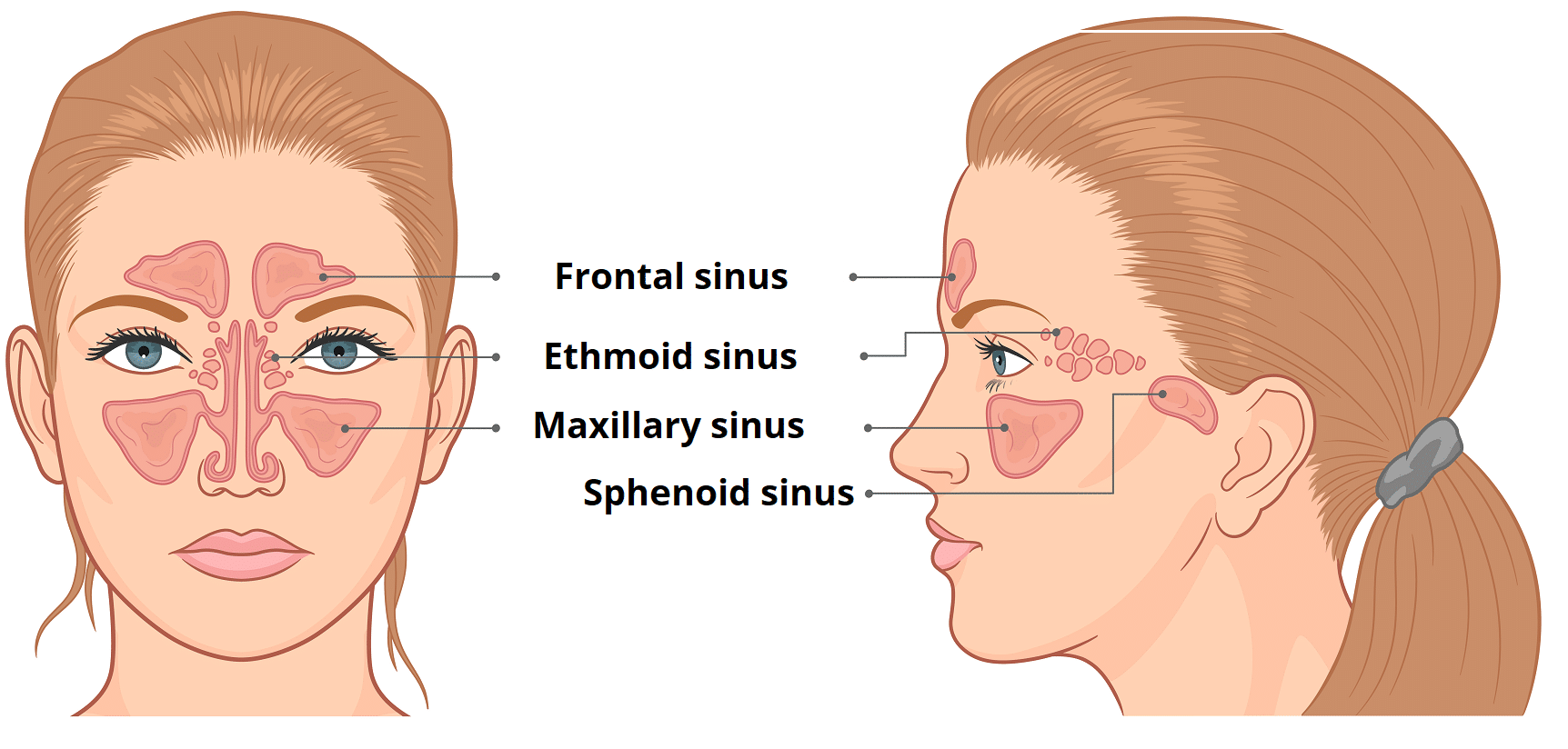


The Paranasal Sinuses Structure Function Teachmeanatomy
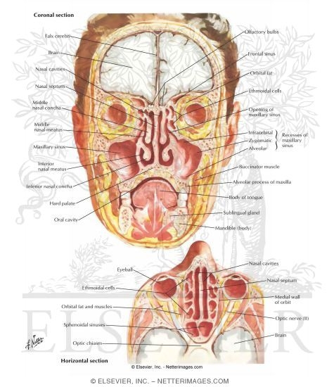


Paranasal Sinuses Nasal Allergy


Paranasal Sinuses Radiology Key


Nasal Cavity And Paranasal Sinus Cancer Miami Cancer Institute Baptist Health South Florida
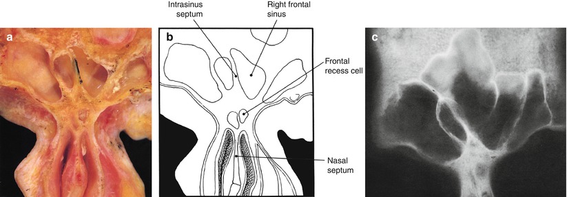


Anatomy Of The Nose And Paranasal Sinuses Springerlink


Human Skull Paranasal Sinus Stock Photos And Images Agefotostock


Nose And Sinuses Amboss


Atlas Of Anatomy Of The Paranasal Sinuses Paranasal Sinuses


Paranasal Sinuses


Figure Anatomy Of The Paranasal Sinuses Spaces Between The Bones Around The Nose Pdq Cancer Information Summaries Ncbi Bookshelf
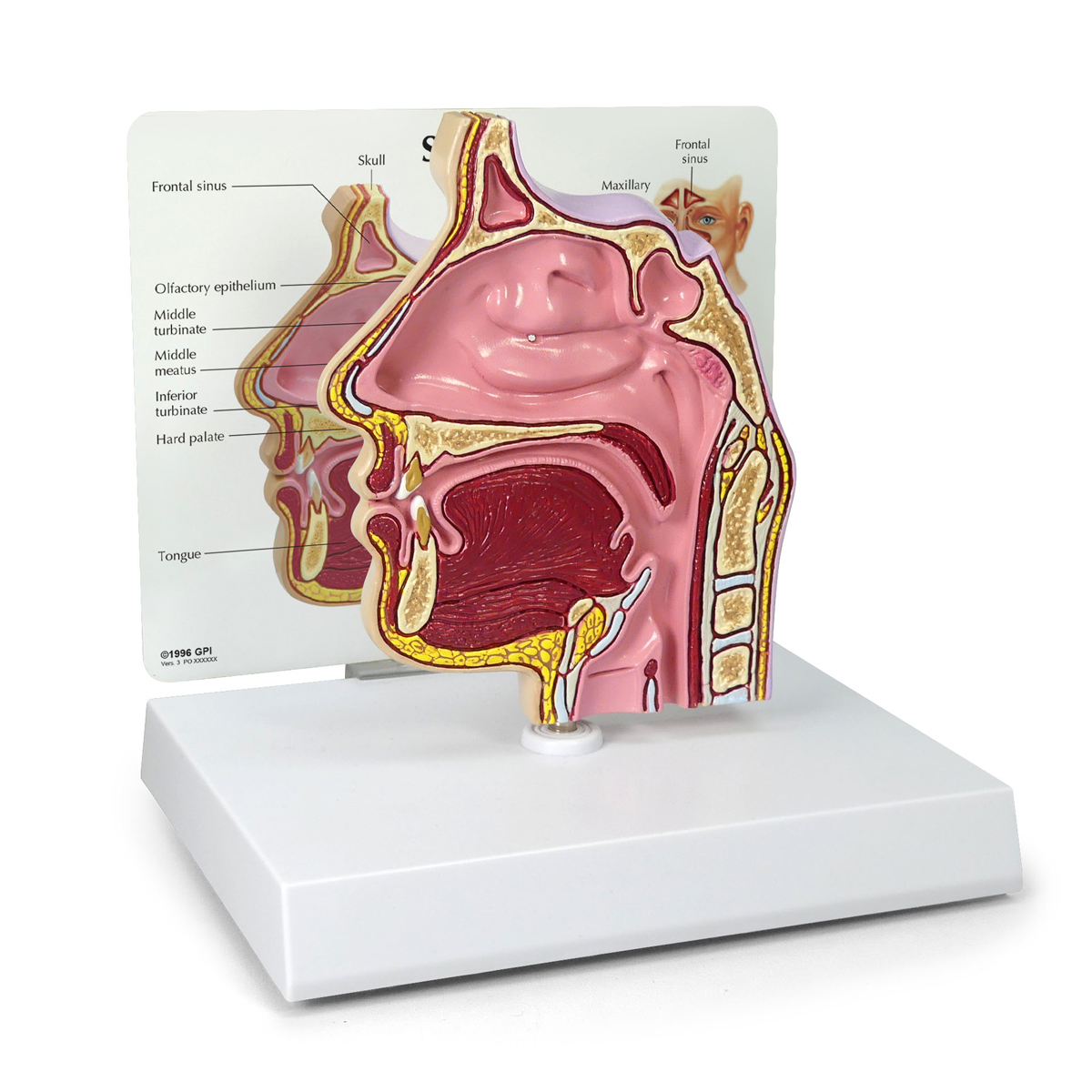


Sinus Cross Section 2850 Anatomical Models Anatomy Teaching Models Cranial Models Head Models


Nose Nasal Cavity Paranasal Sinuses Pharynx Dr Jamila


7 Paranasal Sinus Illustrations Clip Art Istock
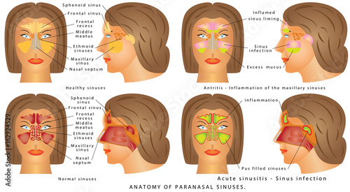


Nasal Sinus Nasal Sinus Human Anatomy Sinus Diagram Anatomy Of The Nose Nasal Cavity Bones Anatomy
:background_color(FFFFFF):format(jpeg)/images/article/en/the-paranasal-sinuses/972PC0nYOzlz7wqSgLmNA_sinus_frontalis_large_u9Vfozc0uUoMtc6KtIaUfw.png)


Paranasal Sinuses Anatomy And Clinical Aspects Kenhub


The Paranasal Sinuses Structure Function Sciencekeys


Diagram Of Paranasal Sinuses Download Scientific Diagram
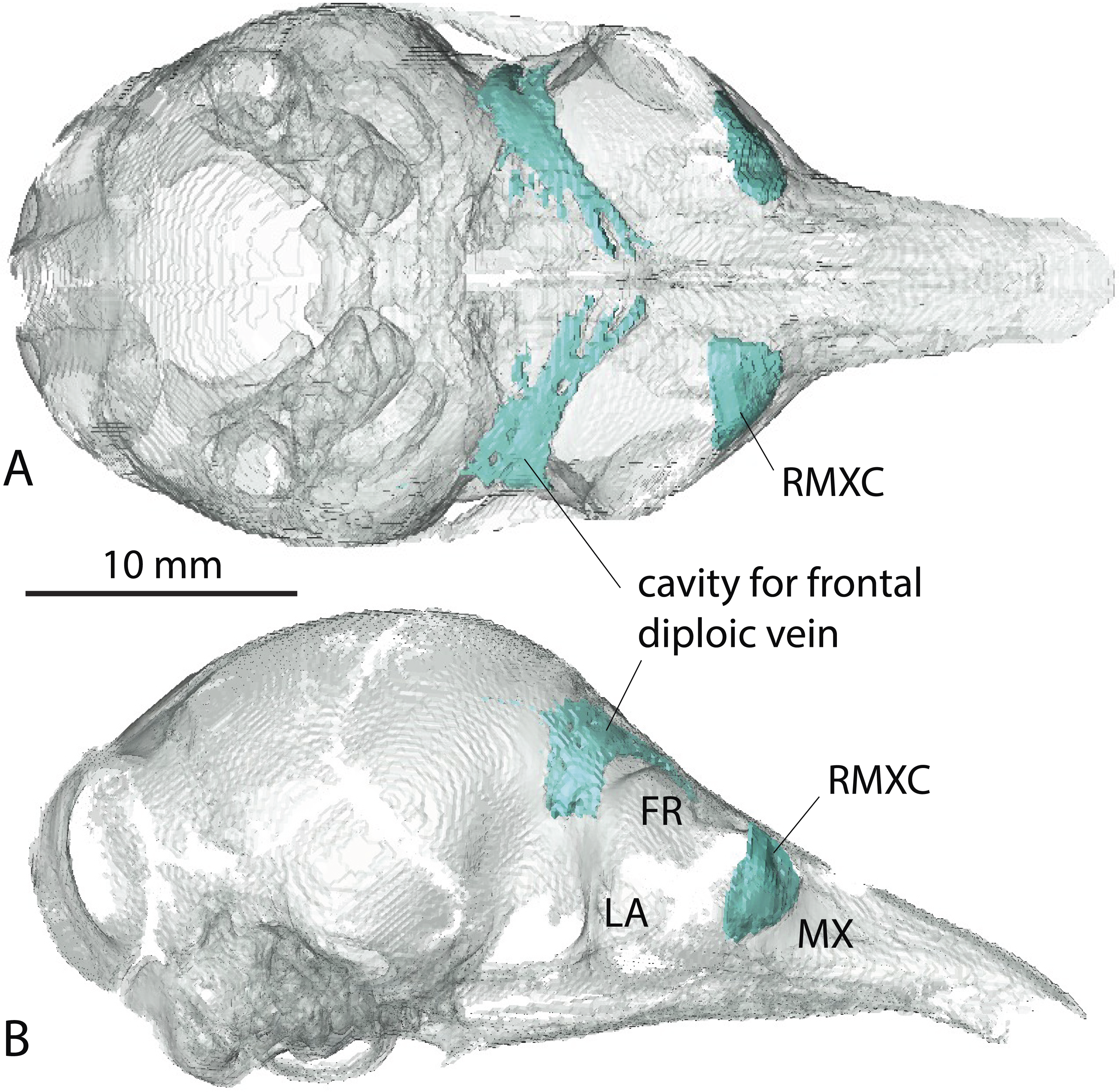


The Hidden Anatomy Of Paranasal Sinuses Reveals Biogeographically Distinct Morphotypes In The Nine Banded Armadillo Dasypus Novemcinctus Peerj


Www Orl Hno Ch Fileadmin User Upload Dokumente Veranstaltungen Sommerschule Sommerschule 18 18 Anatomy Physiology Nose Paranasal Sinuses Handout Pdf
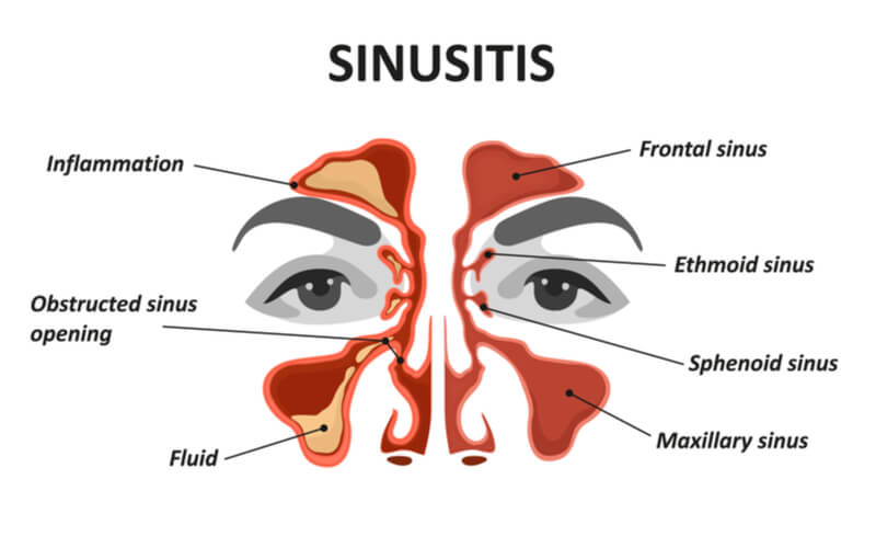


Maxillary Sinus The Definitive Guide Biology Dictionary


Anatomy Of Nose And Paranasal Sinus Ppt Video Online Download
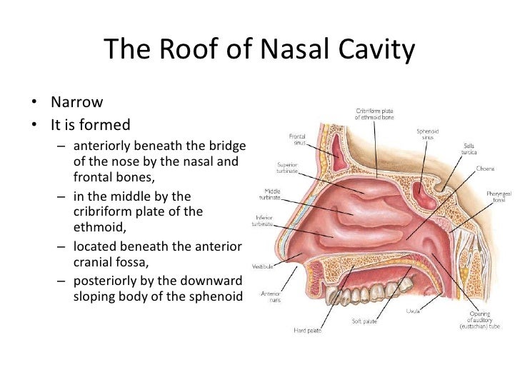


Anatomy Of Nose And Paranasal Sinus
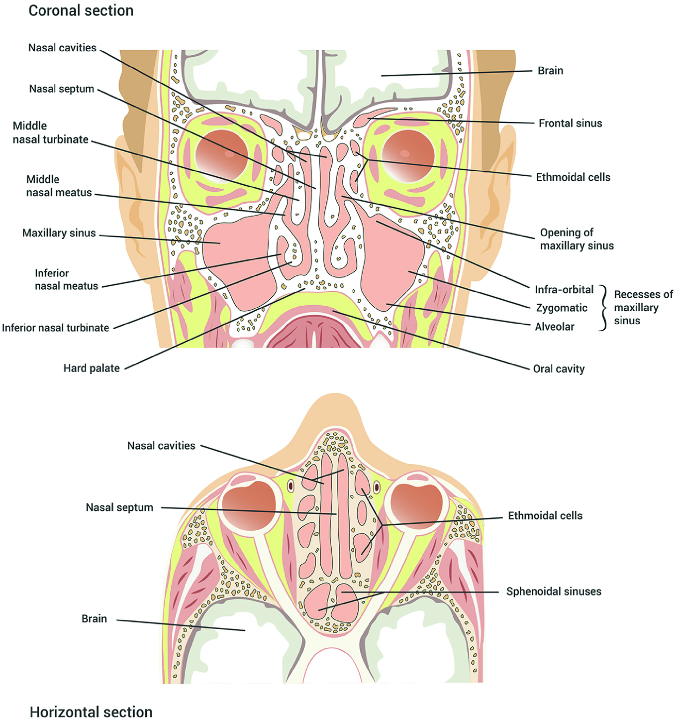


Anatomy And Physiology Of The Human Nose Springerlink


コメント
コメントを投稿